
The parotid gland is the hidden indicator of training quality and here is why.
Invisible glands are good
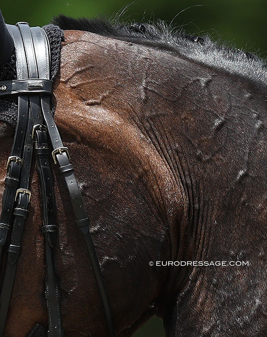
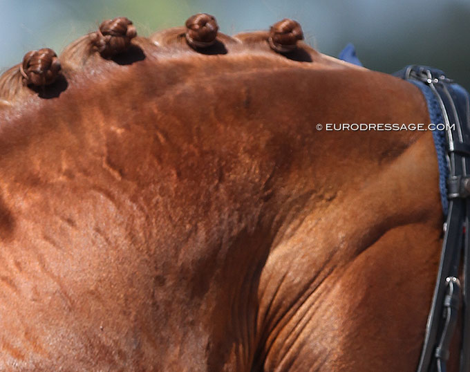
When you don’t see them, it is most likely a good sign. Parotid glands are usually clearly visible on horses that tend to move in a short, tense, and compressed frame. This can be caused by bad training, and some horses may offer this frame themselves – in both cases the horse needs to learn to trust the rider and reach to the bit in a soft and balanced way.
If the frame is relaxed and correct, the parotid glands are normally invisible. If the frame is closed, the parotid gland is pushed out, as seen on the video. The salivary system is sensitive, and it can react to many things. If there are any visible changes they should be checked by a veterinarian to exclude tooth issues, allergic reactions and other problems.
Parotid gland is the most well known
Largest salivary gland, parotid, is easiest to see due to its location. The Parotid gland fills the space between the first cervical vertebra, atlas joint, mandible, and base of the ear.
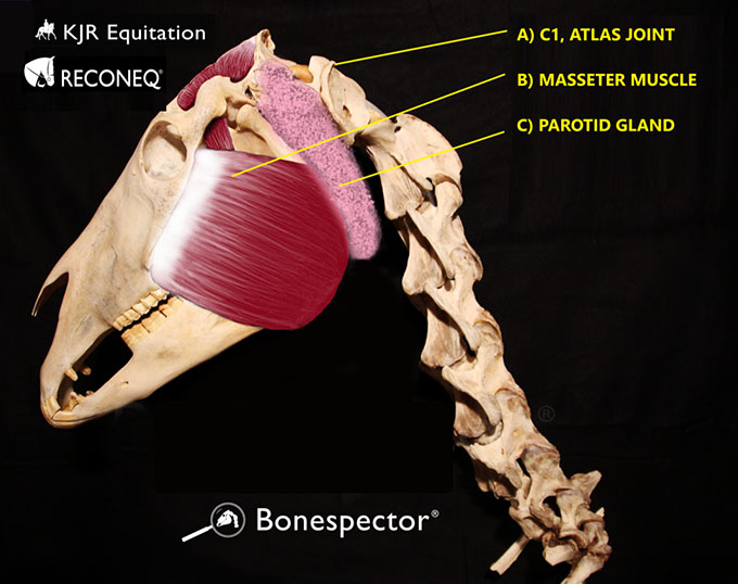
the mandible and the first cervical vertebra
Parotid gland is covered with parotidoauricularis muscle, which runs from the ear to the fascia of the parotid gland. The parotid gland is connected to the mouth via the parotid duct. This duct travels medially behind the mandible, it joins the route of facial artery and vein in front of the masseter muscle, and dives into the mouth near the 2nd and 4th cheek teeth. Descriptions of the location have some variation, and with anatomy there is always some individual variation.
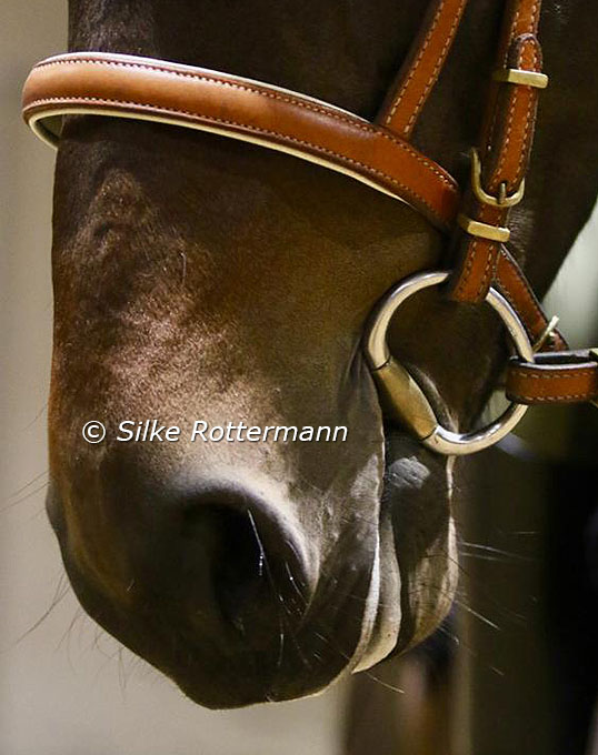
This is good to keep in mind when choosing and fitting a noseband – a heavily padded noseband or special design of anatomical bridle could cause pressure towards this important area and pressure on this area in the lower jaw could cause changes in saliva production.
Mandibular salivary gland hides behind
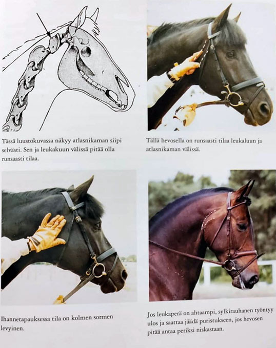
with Kyra' how important it is to recognize
the conformation and its effects
on the frame that is anatomically possible
for the horse (Photos © Jytte Lemkow)
Tightness or swelling in the throat latch area is problematic because the space there is very limited, and the area is very sensitive not only physiologically, but also psychologically due to breathing and swallowing. Also even a light yet constant pressure can be uncomfortable, a feeling of having a lump in one's throat is rather disturbing and it can be caused for many reasons. A constant uncomfortable feeling can possibly rise the sympathetic nervous system activity which can be problematic over the course of time.
Contraction and oedema
Tension leads to lack of gliding between the structures (1,2), which can cause compression of the other structures. Oedema has the same effect.
Oedema can also function kind of the same way, because the fluid in the tissues fills all the space it can possibly find, creating pressure. If oedema is present a long time, it starts to change into fibrosis, which will restrict the function of the area from there on. If the free gliding function between the structures is not working normally, it can cause uncomfortable feeling, tightness, and weird pain symptoms. Oedema can easily become chronic, if there is repetitive tightness in the throat area for example in the daily training.
For this reason the correct and variable frame, the quality of rein contact, and the overall balance and suppleness of the horse are very important ingredients of good training.
The saliva
amount of saliva that forms a soft, thin lipstick,
that is preferably symmetrical.
Lack of space
If the frame is closed, it has several direct effects on the salivary gland function. Closing the frame forces the parotid glands outwards over the mandibular creating significant tension on the superficial and deeper structures, at the same time as the parotid duct gets less or no room to function. Overall tension on the throat area, below the tongue, or on the cheeks causes lack of space that affects the function of the glands. Tension at the throat or tongue affects the breathing directly.
If the contact is good, the frame neutral and in front of the vertical and the tack is bridle is fitting well, and if the rider is having a good communication with the horse, there should be no reason to worry about the glands. If there is still work to be done to learn the horse a correct frame, make sure the work is logical and not depending on who is watching your riding.
In the next part we will look closer at how the rider can help the normal function of the salivary glands.
by Niina Kirjorinne for Eurodressage
Photos © Kirjorinne - Astrid Appels - Silke Rottermann
Sources and further reading:
1. Somatic dysfunction and fascia’s gliding-potential.
Chaitow, L. (2014). Journal of Bodywork and Movement Therapies, 18(1), 1–3.
https://www.bodyworkmovementtherapies.com/article/S1360-8592(13)00201-5/fulltext

and balance of the horse.
Stecco, C., Fede, C., Macchi, V., Porzionato, A., Petrelli, L., Biz, C., De Caro, R. (2018). Clinical Anatomy, 31(5), 667–676.
https://pubmed.ncbi.nlm.nih.gov/29575206/
3. Bruxism-induced parotitis: A retrospective case series analysis
Teymoortash, A., Hoch, S., Weber, D., Wilhelm, T., & Günzel, T. (2018). Bruxism-induced parotitis: A retrospective case series analysis. CRANIO®, 1–7.
https://pubmed.ncbi.nlm.nih.gov/30048228/
4. Dressage with Kyra - book to be ordered through amazon and other major book outlets.
Related Links
Meet Niina Kirjorinne, Classical Dressage Rider and Physiotherapist
Effect of Noseband Positioning to Fascial and Neural Dynamics of the Head
Functionality in Equitation: Skin Damage from Leg Aids
Niina Kirjorinne's training articles on Eurodressage